Address

KARUNA dental MVZ Aurich
Erdbeerweg 52, 26605 Aurich, Germany

Phone: 04941/ 9742030
fax: 04941/ 9742031
info@karuna-zahnarztpraxis.de


KARUNA dental MVZ Weener
Brombeerenweg 3, 26826 Weener, Germany

Phone: 04953/ 9211690
fax: 04953/ 9211691
teamweener@karuna-zahnarztpraxis.de

Opening hours

Monday 08.00 am - 12.00 pm
2.00 pm - 6.00 pm
Tuesday 08.00 am - 12.00 pm
2.00 pm - 6.00 pm
Wednesday 08.00 am - 12.00 pm
2.00 pm - 5.00 pm
Thursday 08.00 am - 12.00 pm
2.00 pm - 6.00 pm
Friday 08.00 am - 12.00 pm
   

Appointments beyond that by appointment.

Journey

Aurich, Germany

Weener (Near Sanddornweg 2) Blackberry Trail 3

Periodontitis

Treatment of sensitive teeth in periodontitis


Depth measurement of gingival pockets


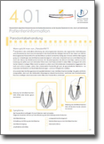
Treatment of deep pockets


Bone loss in periodontitis

Periodontitis (sometimes erroneously called periodontitis) is an inflammation of the tooth bed (periodontium). Like gingivitis, periodontitis is triggered by bacterial plaque. However, the main difference is the bone loss that occurs in periodontitis, which can lead to tooth loss.

In industrialized countries, periodontitis is the most common chronic infectious disease and thus also the most important risk factor for diseases outside the oral cavity. Thus, there is a connection between periodontitis and hardening of the arteries, heart disease, vascular disease as well as stroke and heart attack. The risk of developing periodontitis is increased by smoking, inadequate dental care, improper nutrition and metabolic diseases. Anyone who grinds their teeth at night or breathes with their mouth open also belongs to the risk group.
What many do not know: Periodontitis is quite transmissible. So if the life partner suffers from this disease, the risk of infection is great. Infection from mother to child is also possible.


Commonly, teeth can develop periodontitis unnoticed even with good care. From a simple gingivitis of gingivitis, periodontitis can develop without you noticing clear signs of it. The disease progresses insidiously and usually without pain. In addition to occasional bleeding gums, bad breath, change in tooth position or lengthening and loosened tooth necks may occur.

How does periodontitis develop?
Normally, the gums adhere tightly to the tooth. The periodontium includes the gums (gingiva), the root skin (desmodont) and the jawbone. The root of the tooth is fixed in the jaw with the help of the root skin. The root membrane consists of many thousands of fibers, which firmly connect the tooth with the surrounding jawbone. Due to the close connection, no germs can penetrate. If the plaque is not removed by lack of hygiene, tartar can now form through minerals from the saliva. On its rough surface, in turn, unwanted bacteria can spread. Over time, a so-called biofilm is formed.

In such a biofilm, bacteria are connected in a kind of three-dimensional network. As if mixed with adhesive, they adhere firmly to each other and to the tooth surface. The removal of this viscous mass by simple rinsing is no longer possible. Due to the biofilm, the bacteria are only accessible to the body's own defense and also to drugs such as antibiotics to a limited extent. Protected in this way, they can produce their toxic waste products (toxins), which then drive inflammation of the gums.

The biofilm is thus able to spread further and pushes itself further between the root of the tooth and the gums. The result is the so-called gingival pocket which exposes the tooth more and more.

Due to misinformation in the disturbed cell communication, in addition to the loosening process between the tooth and the tooth bed, the jawbone also breaks down. The end result can be the complete detachment of the tooth from the holding apparatus and thus its loss.

Warning signs of periodontitis can be:
  • Bleeding gums
  • Reduction of gums
  • Touch-sensitive gums
  • Pus discharge when pressed on gums
  • Reddened, swollen gums
  • Bad breath, bad breath
  • Loose or migrating teeth
  • Changes in biting behaviour (bite)
  • Altered prosthesis fit, poor hold of the prosthesis
Since the periodontium inflammation does not initially cause pain, it can remain undetected for a long time. Often it is only recognized in the late stages by occasional bleeding gums. Unfortunately, irreparable bone loss has often occurred here. Regular check-ups with us help to detect periodontitis as early as possible and to avoid major consequential damage.

Course of treatment
In any case, it is very important to treat periodontitis in time. Otherwise, there is a risk of premature tooth loss. First, plaque and tartar are removed. Because only when the tooth surface is smooth, the gums can put on again and the gingival pocket close again permanently. It is also possible to determine the bacteria present there by taking a swab from the gingival pocket. It may be useful to use an antibiotic for support.

Then the bacteria are removed from the gingival pockets With a very fine scraper, the curette, the dentist grates the fossilized plaque from the root surfaces in the gingival pockets. If the jawbone has already shrunk considerably, it can be rebuilt if necessary. (see bone augmentation)

If you have been diagnosed with periodontitis, you should have the depth of your gingival pockets checked regularly. The dentist compares the result with the previous examinations. A one-time check-up and treatment is not enough, otherwise you risk that periodontitis will continue to progress unnoticed.

Word meanings:

Periodontitis   - inflammation of the gums or Periodontium
Periodontium - or periodontum - the periodontium 
Periodontitis  - loss of the gums or periodontium without inflammation
further information about periodontitis
pdf
Parodontitis-10-Fragen.pdf pdf Parodontitis-c.pdf pdf Parodontitistherapie-bzaek.pdf pdf Parodontitis-sso.pdf pdf Parodontitis-PSI-Parodontale-Screening-Index-zur-Früherkennung.pdf
pdf
Parodontitis-und-Herzerkrankungen-c.pdf pdf Parodontitis-Volkskrankheit-prodente.pdf pdf Parondontitis-prodente.pdf pdf Parodontitis-Vorbeugung-und-Therapie-dgp.pdf pdf Parodontitis-blzk.pdf
pdf
Parodontitis-die-unbekannte-Volkskrankheit.pdf pdf Parodontalbehandlung-bzaek.pdf pdf   pdf   pdf  

Ringing in the ears

The sense of hearing and balance are located in the bone directly behind the temporomandibular joint. Therefore, in  20% of all tinnitus patients, the teeth and the temporomandibular joint as well as a tense jaw muscles are the cause of their complaints. There is only a few millimeters between the temporomandibular joint and the inner ear. Those who tend to grind or press their teeth consciously or unconsciously due to stress, worry or high workload tense their masticatory muscles and thus overload their temporomandibular joint. Especially teeth grinding, which affects every fifth adult, not only grinds off the enamel, but also increases the risk of developing tinnitus. Due to the constant overloading of the temporomandibular joint, pressure is also exerted on the fine hair cells of the inner ear. These are thereby damaged, similar to a sudden hearing loss. This is also an explanation for the frequent co-occurrence of stress and tinnitus.

Another trigger for ear noises can be a malocclusion, a displacement of the upper and lower jaw. Here, teething splints made of plastic that are worn at night help. Those who regularly "chew through" their problems in their sleep should not necessarily rely only on the plastic rail. With meditation, yoga and a "decelerated" schedule, you should consistently treat yourself to more relaxation.

Oral Care

further information about oral care for seniors
pdf
Mundpflege-Senioren-c
pdf Prophylaxe-fuer-Senioren
pdf   pdf

Oral hygiene in young children

further information about oral hygiene
pdf
Schwangerschaft-c
pdf
pdf   pdf

Oral mucosal diseases

Most often, a disease of the oral mucosa is harmless. However, any change in the oral mucosa should be checked so that it can be determined whether therapy is necessary or not. Oral mucosal diseases are usually not noticed because they are often absolutely asymptomatic. Since many skin and general diseases are noticeable as changes in the oral mucosa, the dentist is of great importance in the semi-annual routine check of your teeth.
In very rare cases, it can lead to a degeneration of oral mucosal cells. If malignant changes are detected early, action can be taken quickly and greater damage can be avoided.

Some typical diseases of the oral mucosa are:

Aphthae

Aphthae are white, painful vesicles in the oral cavity that occur acutely and are usually lentil-sized. These can be caused by immunodeficiency, stress, injuries to the oral mucosa or hormone fluctuations. They interfere with speaking, chewing and swallowing. With the help of mouthwashes and adhesive ointments, they heal without scarring within 10 to 14 days.

Stomatitis

Symptoms of stomatitis are the reddened and inflammatorily altered oral mucosa, as well as bad breath, loss of appetite and easily triggered oral mucosal bleeding. In addition to bacterial infections, there are other causes of inflammation of the oral mucosa, such as:

  • inadequate dental/oral / prosthesis care
  • Burns caused by food or drinks that are too hot
  • Nicotine/alcohol abuse
  • dry oral mucosa
  • Vitamin deficiency
  • Irritation and injuries of the oral mucosa
  • weakened immune system

Depending on the cause, drug treatment may be necessary.

Oral thrush

This fungal infection, caused by a yeast fungus, can be recognized by cream-colored, yellow-white or red spots on the oral mucosa that can be wiped off. The tissue under these areas can hurt. Also, patients with thrush infestation may have bad breath. With the appropriate antifungal agents (antifungals) oral thrush can be treated very well.

Leukoplakia

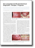
This change in the oral mucosa is considered a precursor to cancer. The mucous membrane is thick and white and can not be wiped off. The cause of this is usually tobacco consumption or chewing on the cheek. If your dentist detects leukoplakia, he will remove tissue from you to determine whether it is a benign or malignant tissue change.

Oral cancer

Cancer is a malignant neoplasm of tissue. In the interior of the mouth, malignant tissue changes occur in various places in oral cancer. These tissue changes usually need to be removed and/or treated with radiotherapy and/or chemotherapy. Early detection of the disease is therefore very important for successful treatment.

further information on the topic of oral mucosal diseases
pdf
Mundschleimhautveraenderungen-c
pdf Leukoplakie pdf Mundhoehlenkrebs-zzq            
Mundkrebs-krebshilfe pdf Mundschleimhauterkrankungen-uni-leipzig pdf Mundschleimhauterkrankungen-zwp